Zhang Kai, associate researcher of the Beijing Synchrotron Radiation Facility, Institute of High Energy Physics, Chinese Academy of Sciences, and other domestic and foreign research teams have made progress in quantitative research on the decline mechanism of chemical-mechanical interactions in lithium-ion batteries using synchrotron multi-scale imaging technology The research results were recently published in the journal Advanced Energy Materials.
With the advancement of science and technology, there is an increasing urgent demand for energy in all walks of life. At the same time, people are also facing the energy crisis and the environmental problems caused by traditional energy. Therefore, people are constantly looking for new energy materials that are inexpensive, environmentally friendly, excellent in performance, and safe. Among them, high-efficiency energy storage devices represented by lithium batteries have become one of the important directions of new energy research. As a typical composite multiscale heterogeneous system, although the basic ion migration of a lithium-ion battery system occurs at the atomic scale, the most important device performance indicators, such as energy density, high-speed charge / discharge capability, and material life, all depend on multiple The combined effect of different scales. Therefore, how to implement multi-scale imaging methods in order to study the three-dimensional morphology structure and crystal equivalent information of the composite electrode in the range of micro, mesoscopic and macro scales, to develop high efficiency and high stability for understanding the migration of ions and charges in the device Battery material is critical.
In recent years, thanks to the use of synchrotron radiation light source technology and the development of imaging theory and optical component manufacturing and processing technology, synchrotron radiation X-ray microscopic imaging technology can realize multi-scale three-dimensional nondestructive structure imaging from nanometer resolution to micrometer resolution. In addition, the synchrotron radiation X-ray spectroscopy imaging technology combined with this technology and near-edge absorption spectroscopy technology can non-destructively reconstruct the three-dimensional morphology, element distribution and valence state heterogeneity of electrode materials in lithium batteries under in-situ environment. information. This makes synchrotron radiation X-ray micro-imaging technology the most direct and effective method for studying the correlation between the multi-scale three-dimensional topography and performance of composite electrode materials, and opens a new window for understanding the microscopic world of electrode materials.
Kai Kai and others and domestic and foreign cooperation teams have used synchrotron radiation imaging technology to quantitatively analyze the chemical-mechanical interactions in lithium battery materials at mesoscale (secondary particle) and macroscale (electrode level), and related research results have been published recently. In Advanced Energy Materials magazine (Adv. Energy Mater. 2019, 1900674). Yang Yang (European Synchrotron Radiation Light Source), Xu Rong (Purdue University), Zhang Kai (Beijing Synchrotron Radiation Light Source) are co-first authors of the paper, Zhao Kejie (Purdue University), Lin Feng (Virginia Institute of Technology) and Liu Yijin (Stanford Synchronization) Radiation source) is the co-corresponding author of the paper.
They first studied the nickel-rich LiNi1-x-yMnxCoyO2 (NMC) secondary particles with a diameter of 10 microns using nano-resolution (30 nanometers) spectroscopy imaging technology, and found that lithium battery particles will generate a large number of cracks during charge and discharge, including The initial cracks and mature cracks are developed, as shown by the green arrows and red arrows in Figure 1 (i). Further X-ray spectroscopy imaging revealed that the mature crack surface showed a higher oxidation state, as shown in Figure 1 (j). This is caused by the penetration of the liquid electrolyte and the precipitation of lithium ions at the new solid-liquid interface. However, due to the lack of liquid electrolyte infiltration in the initial cracks, the nickel oxidation valence state of these regions has not changed significantly compared with other bulk phase regions of the entire battery particles, as shown in the energy distribution diagram of k-edge of Ni in (k) Ni Show.
On this basis, in order to study the influence of the development and change of internal cracks of battery particles on the performance of lithium batteries, researchers used X-ray phase contrast imaging technology to perform chemical-mechanical interactions of thousands of battery particles in the entire electrode material. Imaging experiment research systematically analyzes the imbalance of local conductivity and ion conductivity reflected by the non-uniform damage of battery particles on mesoscopic and macro scales. The study found that under fast charging conditions, a significant reconstruction of the entire electrode surface occurred (see Figure 2c.d), and the top of the electrode (near the separator) experienced a more severe local phase transition from a layered structure to spinel and The mixture of rock salt structure, most of the three-dimensional morphological structure of the battery particles have been severely damaged; and the three-dimensional morphological structure of the particles at the bottom of the electrode sheet has been relatively well preserved, as shown in Figure 2d.f. The above observations indicate that there is an uneven reaction on the electrode scale, which is very obvious in the depth direction and the lateral direction.
This work provides important information for researchers to understand the performance decay mechanism of lithium battery cathode materials, which can help people establish the relationship between the macroscopic properties and microscopic material structures of battery materials. This information not only helps material scientists to further improve the properties of battery materials from an application perspective, but also has important value for basic scientific research on battery material failure mechanisms. The multi-scale imaging technology used in the study has important reference significance for many subject areas of cutting-edge research using large scientific devices.
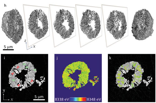
Fig.1 Structural defect distribution of NMC622 battery particles at the nanometer scale. (h) Structure distribution information of single NMC622 battery particles obtained by X-ray nano-resolution spectroscopy imaging at different faults (ZX direction) (i) Tomographic slice images of NMC622 battery (YX direction), red arrows show mature cracks , The green arrow shows the initial crack. (J) is the distribution image of the K-edge energy of Ni on the tomographic image (i) of the NMC622 battery (i) is the distribution of the K-edge energy of Ni near the mature crack.
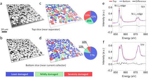
Fig. 2 The morphology of the top slice and the bottom slice after the NMC622 electrode material undergoes 10 rapid charge-discharge cycles (charging rate 5C). (a) Top tomographic image of NMC622 electrode material. (c) NMC622 electrode material top tomographic slice image after image segmentation. (b) The tomographic image of the bottom of the NMC622 electrode material. (d) NMC622 electrode material bottom tomographic slice image after image segmentation. (e) (f) Soft X-ray absorption spectrum test results obtained using TEY and TFY modes for the bottom (blue curve) and top (red curve) classification of NMC622 electrode materials.
Rubber expansion joint system comprises of No.8 standard models designed to accommodate movement up to 330mm by shear deformation of the elastomer between the steel components. Each model of our rubber plate-type expansion joint incorporates steel angles designed to be bolted to the structural deck and a steel bridging plate system which spans the open joint gap. The elastomer case is highly resistant to oils, solvent spillage and the trafficked surface includes an anti-skid pattern for safety having a rubber to rubber coefficient of static friction of 0.69. Each model of bridge deck Rubber Expansion Joint is specifically designed to accommodate horizontal and skew movement and will also accommodate vertical movement due to rotation of up to 6mm.J.S Brown can supply many kinds of movements rubber expansion joints such as 30mm movement rubber expansion joint,50mm movement rubber expansion joint and so on.We have exported to many countries.

Rubber Expansion Joint
Rubber Expansion Joint,Single Ball Rubber Expansion Joint ,Flexible Rubber Expansion Joint,Customized Rubber Expansion Joint
J.S.Brown New Material Technology Co., Ltd. , https://www.profile-steel.com